
#How to merge two images in imagej software online professional
To create a professional looking publication one must have consistent format within and between figures. A major publication can contain over 200 image panels. Microscopy intensive scientific manuscripts often include figures with large numbers of fluorescent microscope images often split by channel into separate panels. The software is free, open source and can be installed easily. Exported objects and text are editable in their target software, making them suitable for sharing with collaborators. Export was successfully tested for each file format and object type. After a user has saved time by creating their work in QuickFigures, the figures can be exported to a variety of formats including PowerPoint, PDF, SVG, PNG, TIFF and Adobe Illustrator. Therefore, QuickFigures is an advantageous alternative to traditional methods of constructing scientific figures.

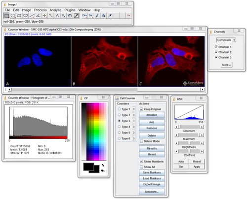
QuickFigures had the most extensive set of features. The toolsets were also compared by checking each software against a list of features. QuickFigures significantly reduced the amount of time required to create a figure. QuickFigures was compared to previous tools by measuring the amount of time needed for a user to create a figure using each software (QuickFigures, OMERO.figure. Those features include tools that automatically create split channel figures from a region of interest (“Quick Figure” button and “Inset Tool”), layouts that make it easy to rearrange panels, multiple tools to align objects, and “Figure Format” menu options that help a user ensure that large numbers of figures have consistent appearance. QuickFigures includes many helpful features that streamline the process of creating, aligning, and editing scientific figures. In order to save time, I have created a toolset and ImageJ Plugin called QuickFigures. Assembling and editing these figures with even spacing, consistent font, text position, accurate scale bars, and other features can be tedious and time consuming. Similar layouts of panels are used when displaying other microscopy images, electron micrographs, photographs, and other images. Publications involving fluorescent microscopy images generally contain many panels with split channels, merged images, scale bars and label text.


 0 kommentar(er)
